2nd Intercostal Space
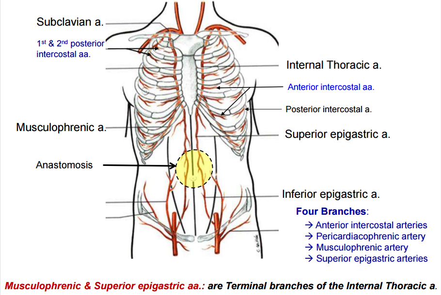
Intercostal arteries and intercostal veins.
2nd intercostal space. Der interkostalraum ist der raum zwischen zwei benachbarten rippen. Since there are 12 ribs on each side there are 11 intercostal spaces each numbered for the rib superior to it. At the back of. Dimitrios mytilinaios md phd last reviewed.
They pass forward in the intercostal spaces below the intercostal vessels. Im interkostalraum befinden sich folgende strukturen. May 15 2020 the eleven paired intercostal spaces contain the intercostal muscles nerves arteries veins and investing fascia. 2 nd intercostal space starts from inferior portion of 2 nd rib and ends at superior portion of 3 rd rib.
Every space consists of intercostal muscles and neurovascular bundle of intercostal plane. On each side 11 intercostal spaces are present. The 2nd rib is continuous with the sternal angle. The intercostal spaces are the spaces amongst the two bordering ribs and their costal cartilages.
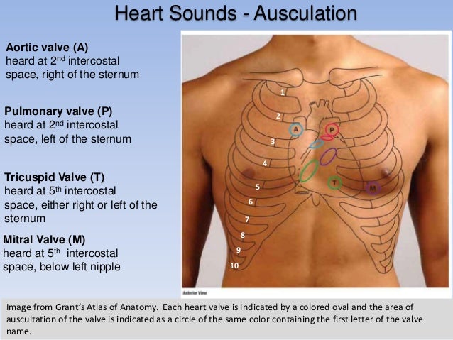
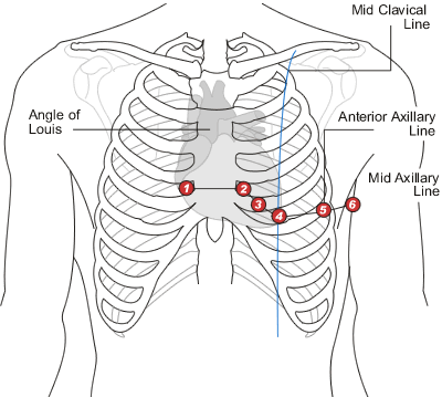
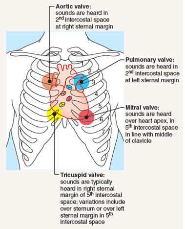
The intercostal space ics is the anatomic space between two ribs lat. The angle of louis also marks the site of bifurcation of the trachea into the right and left main bronchi and corresponds with the upper border of the atria of the heart. If the timing of extra heart sounds and murmurs is confusing at the lower sternal edge or apex as it often is in patients with fast heart rhythms the clinician can return the. At the second left intercostal space s 2 is generally louder shorter and sharper than s 1 s 2 has more high frequency energy than s 1 which is why dup a snappier sound than lub is used to characterize s 2.
Die reihenfolge der interkostalen leitungsbahnen laesst sich mit van gut merken zu oberst vene zu unterst nerv. Reference lines help pinpoint findings vertically. The intercostal space ics is the space in between the two adjacent ribsthere are 11 intercostal spaces on each side each intercostal space is numbered for the rib superior to it. Matthew grouthamel md reviewer.
The structures in each intercostal space are supplied by a single large posterior intercostal artery and 2 smaller anterior intercostal arteries. Bordered by the rib above and below the deep fascia of the thorax superficially and the endothoracic fascia and pleura internally the intercostal. The anterior divisions of the second third fourth fifth and sixth thoracic nerves and the small branch from the first thoracic are confined to the walls of the thorax and are named thoracic intercostal nerves. In the first and second intercostal spaces a large posterior artery arises from the superior intercostal artery a branch from the costocervical artery.
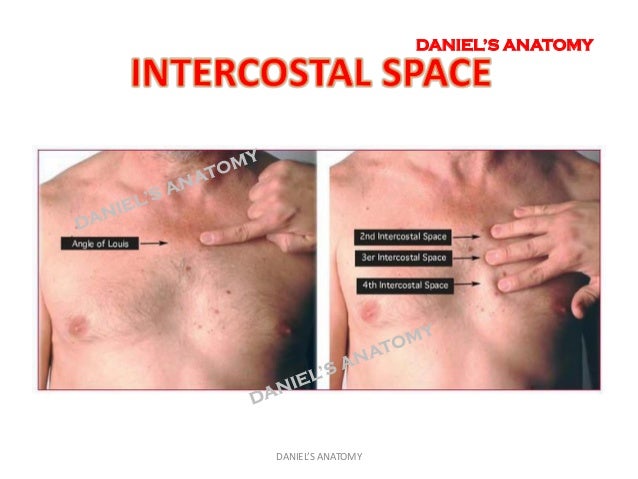
Figure 4 5 shows the location of the second intercostal space. Structures in intercostal space. Several kinds of intercostal muscle.
